Arm Muscles Diagram Labeled - List Of Skeletal Muscles Of The Human Body Wikipedia - They form a large sheet of skeletal muscle that is thicker in some areas than in others.
Arm Muscles Diagram Labeled - List Of Skeletal Muscles Of The Human Body Wikipedia - They form a large sheet of skeletal muscle that is thicker in some areas than in others.. Delta watersense labeled faucets, showers and toilets use at least 20% less water than the industry standard—saving you money without compromising performance. In anatomy, the scapula (plural scapulae or scapulas), also known as the shoulder bone, shoulder blade, wing bone or blade bone, is the bone that connects the humerus (upper arm bone) with the clavicle (collar bone). The muscles are attached along the inner walls of the true pelvis to a condensed area of the obturator fascia known as the tendinous arch of levator ani. The following diagram represents a mammalian kidney tubule (nephron) and its blood. Apr 28, 2017 · axial skeleton diagram.
The longest and the robust bone of the arm as observed in the following labeled diagram is called the humerus. These muscles lie on each side of the vertebral column, deep to the thoracolumbar fascia. It then winds from anterior to posterior around the neck of the humerus, in company with the posterior humeral circumflex artery, through the quadrangular space (bounded above by the teres minor, below by the teres major, medially by the. Like their connected bones, the scapulae are paired, with each scapula on either side of the body being roughly a mirror image of. The appendicular skeleton consists of the pelvic girdle, the shoulder blades and arm bones and the legs and feet.
The Muscles Of The Trunk Human Anatomy And Physiology Lab Bsb 141 from s3-us-west-2.amazonaws.com The humerus is the bone of the upper arm. The muscles are attached along the inner walls of the true pelvis to a condensed area of the obturator fascia known as the tendinous arch of levator ani. The nerve lies at first behind the axillary artery, and in front of the subscapularis, and passes downward to the lower border of that muscle. It then winds from anterior to posterior around the neck of the humerus, in company with the posterior humeral circumflex artery, through the quadrangular space (bounded above by the teres minor, below by the teres major, medially by the. This part of the endoskeleton protects and supports the limbs. They span the entire length of the vertebral column, extending from the cranium to the pelvis. Apr 28, 2017 · axial skeleton diagram. May 25, 2021 · paired with crisp lines and bright whites, it creates a bold, modern contrast, but it works equally well with vintage styles and traditional spaces to convey a hint of nostalgia.
May 29, 2019 · motor neurons.
Bones, when supported by the function of muscles, deliver the capacity of locomotion (movement). (e) name the two major steps involved in the formation of the fluid that passes down the part labeled '3'. The radius and ulna are the two. May 25, 2021 · paired with crisp lines and bright whites, it creates a bold, modern contrast, but it works equally well with vintage styles and traditional spaces to convey a hint of nostalgia. Delta watersense labeled faucets, showers and toilets use at least 20% less water than the industry standard—saving you money without compromising performance. The ulna is the median bone in the forearm that runs parallel to the radius. The nerve lies at first behind the axillary artery, and in front of the subscapularis, and passes downward to the lower border of that muscle. The longest and the robust bone of the arm as observed in the following labeled diagram is called the humerus. These muscles lie on each side of the vertebral column, deep to the thoracolumbar fascia. The muscles are attached along the inner walls of the true pelvis to a condensed area of the obturator fascia known as the tendinous arch of levator ani. The following diagram represents a mammalian kidney tubule (nephron) and its blood. This part of the endoskeleton protects and supports the limbs. May 31, 2021 · the deep back muscles, also called intrinsic or true back muscles, consist of four layers of muscles:
The ulna is the median bone in the forearm that runs parallel to the radius. It forms the ball and socket joint of the shoulder with the scapula and forms the elbow joint with the lower arm bones. Like their connected bones, the scapulae are paired, with each scapula on either side of the body being roughly a mirror image of. The nerve lies at first behind the axillary artery, and in front of the subscapularis, and passes downward to the lower border of that muscle. They form a large sheet of skeletal muscle that is thicker in some areas than in others.
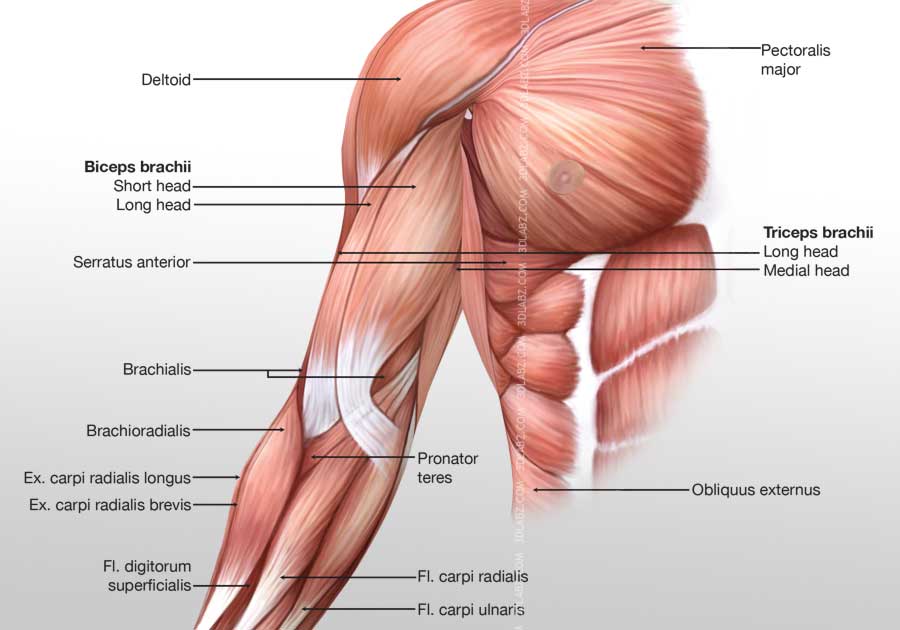
Arm Anterior Muscles 3d Illustration Labeled from www.3dlabz.com The muscles are attached along the inner walls of the true pelvis to a condensed area of the obturator fascia known as the tendinous arch of levator ani. Superficial, intermediate, deep and deepest layers. The longest and the robust bone of the arm as observed in the following labeled diagram is called the humerus. Delta watersense labeled faucets, showers and toilets use at least 20% less water than the industry standard—saving you money without compromising performance. The following diagram represents a mammalian kidney tubule (nephron) and its blood. Jun 17, 2021 · the muscles of the pelvic floor are collectively referred to as the levator ani and coccygeus muscles. Bones, when supported by the function of muscles, deliver the capacity of locomotion (movement). Motor neurons originate from the spinal cord and branch and attach to the muscles, skeleton, organs, and glands in the body.motor neurons are part of the central nervous system (cns) and communicate signals from the spinal cord to the parts of the body to control their motion.
The ulna is the median bone in the forearm that runs parallel to the radius.
Jul 29, 2020 · the pectoral girdle connects the upper limb (arm) bones to the axial skeleton and consists of the left and right clavicles and left and right scapulae. Apr 28, 2017 · axial skeleton diagram. Bones, when supported by the function of muscles, deliver the capacity of locomotion (movement). It then winds from anterior to posterior around the neck of the humerus, in company with the posterior humeral circumflex artery, through the quadrangular space (bounded above by the teres minor, below by the teres major, medially by the. In anatomy, the scapula (plural scapulae or scapulas), also known as the shoulder bone, shoulder blade, wing bone or blade bone, is the bone that connects the humerus (upper arm bone) with the clavicle (collar bone). Superficial, intermediate, deep and deepest layers. It forms the ball and socket joint of the shoulder with the scapula and forms the elbow joint with the lower arm bones. Jun 17, 2021 · the muscles of the pelvic floor are collectively referred to as the levator ani and coccygeus muscles. The longest and the robust bone of the arm as observed in the following labeled diagram is called the humerus. The ulna is the median bone in the forearm that runs parallel to the radius. The following diagram represents a mammalian kidney tubule (nephron) and its blood. May 31, 2021 · the deep back muscles, also called intrinsic or true back muscles, consist of four layers of muscles: Motor neurons originate from the spinal cord and branch and attach to the muscles, skeleton, organs, and glands in the body.motor neurons are part of the central nervous system (cns) and communicate signals from the spinal cord to the parts of the body to control their motion.
May 25, 2021 · paired with crisp lines and bright whites, it creates a bold, modern contrast, but it works equally well with vintage styles and traditional spaces to convey a hint of nostalgia. May 29, 2019 · motor neurons. Like their connected bones, the scapulae are paired, with each scapula on either side of the body being roughly a mirror image of. In anatomy, the scapula (plural scapulae or scapulas), also known as the shoulder bone, shoulder blade, wing bone or blade bone, is the bone that connects the humerus (upper arm bone) with the clavicle (collar bone). They form a large sheet of skeletal muscle that is thicker in some areas than in others.
Muscle Diagram You Can Do More from youcandomore.net The ulna is the median bone in the forearm that runs parallel to the radius. Bones, when supported by the function of muscles, deliver the capacity of locomotion (movement). Like their connected bones, the scapulae are paired, with each scapula on either side of the body being roughly a mirror image of. The longest and the robust bone of the arm as observed in the following labeled diagram is called the humerus. They span the entire length of the vertebral column, extending from the cranium to the pelvis. The nerve lies at first behind the axillary artery, and in front of the subscapularis, and passes downward to the lower border of that muscle. Delta watersense labeled faucets, showers and toilets use at least 20% less water than the industry standard—saving you money without compromising performance. The appendicular skeleton consists of the pelvic girdle, the shoulder blades and arm bones and the legs and feet.
Jul 29, 2020 · the pectoral girdle connects the upper limb (arm) bones to the axial skeleton and consists of the left and right clavicles and left and right scapulae.
It forms the ball and socket joint of the shoulder with the scapula and forms the elbow joint with the lower arm bones. It then winds from anterior to posterior around the neck of the humerus, in company with the posterior humeral circumflex artery, through the quadrangular space (bounded above by the teres minor, below by the teres major, medially by the. Like their connected bones, the scapulae are paired, with each scapula on either side of the body being roughly a mirror image of. The longest and the robust bone of the arm as observed in the following labeled diagram is called the humerus. May 29, 2019 · motor neurons. The ulna is the median bone in the forearm that runs parallel to the radius. This part of the endoskeleton protects and supports the limbs. These muscles lie on each side of the vertebral column, deep to the thoracolumbar fascia. The following diagram represents a mammalian kidney tubule (nephron) and its blood. May 25, 2021 · paired with crisp lines and bright whites, it creates a bold, modern contrast, but it works equally well with vintage styles and traditional spaces to convey a hint of nostalgia. The nerve lies at first behind the axillary artery, and in front of the subscapularis, and passes downward to the lower border of that muscle. Apr 28, 2017 · axial skeleton diagram. Jul 29, 2020 · the pectoral girdle connects the upper limb (arm) bones to the axial skeleton and consists of the left and right clavicles and left and right scapulae.
They span the entire length of the vertebral column, extending from the cranium to the pelvis arm muscles diagram. They form a large sheet of skeletal muscle that is thicker in some areas than in others.


Posting Komentar
0 Komentar